


Abscesses develop when bacteria enter a part of the body. It is the body’s natural defences to try and ‘wall off’ infection to stop it spreading elsewhere within the body. This can lead to problems when the abscess is located within the region of the eye, since the location is hard to successfully operate on, and the case is frequently difficult to cure.
Abscesses are a common occurrence in rabbits, and form when bacteria enter the skin or a body cavity, and sets up an infection. This causes a pocket of infection to form, characterised by an accumulation of pus.
Unlike dog and cat pus (exudate), rabbit exudate is thick and semi-solid, much like toothpaste, making it difficult to flush. This causes a high recurrence rate, since only a small amount of exudate needs to be left inside the abscess cavity for it to return.
Abscesses that are located around the face are often due to dental disease, which can also manifest as ocular problems, since the upper tooth roots can overgrow and impinge upon the eye socket and the nasolacrimal tear duct.
Abscesses that develop behind the eye are known as retrobulbar abscesses. They are very well encapsulated and because of their location they are very difficult to reach and drain.
Dental disease is often a predisposing factor since the roots of the upper cheek teeth can impinge upon the eyeball, and in extreme cases penetrate through it. The food passes from the mouth to the socket of the root developing infection that eventually becomes an abscess.
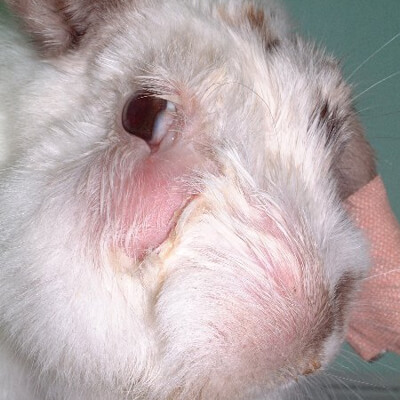
The most common signs of a retrobulbar abscess are bulging of the eye, protrusion of the third eyelid across the eye and inability to close the eyelids completely. Abnormal ocular or oculonasal discharge can also develop as a consequence of the eye irritation and the rabbit may show signs of discomfort such as excessive blinking and grinding teeth.
In chronic cases, rabbits can continue eating and drinking normally, but often the rabbit will deteriorate as the pain escalates or the dental disease progresses and eventually will stop eating and passing faecal pellets.
There are many investigations that are possible to perform in order to make a diagnosis of retrobulbar abscess; the first is the a physical examination. Often, in moderate to severe cases, it is possible to make a presumptive diagnosis after having examined the affected eye and the mouth for the presence of dental disease.
However, in order to confirm the diagnosis, radiographic and ultrasonographic examinations are generally the tests of choice. Radiography (x-rays) of the skull is generally performed under general anaesthesia. X-rays will not only show abnormalities behind the eye, but will also give the opportunity to confirm or rule out dental disease.
Ultrasonography (scan) of the eye can be performed when the animal is conscious or under light sedation depending on the nature of the animal. However, this test is limited to the evaluation of the eye and the retrobulbar space.
Ideally a computed tomography (CT) or magnetic resonance imaging (MRI) scan should be taken to assess the extent of the abscess, the infection and the tooth roots, but often this is not possible due to financial constraints.
It is also posssible to aspirate material from the abscess and test it for bacteria in order to make a diagnosis of infection rather than tumour, and also to choose the best antibiotics as treatment.
Unfortunately, retrobulbar abscesses are very difficult to treat due to the thick exudate that is difficult to drain and due to the location just behind the eye.
Medical treatment is generally unrewarding and surgical removal of the eye (enucleation) is usually necessary. Surgery allows complete removal of the abscess and a thorough flushing of the cavity to ensure that none of the exudate is left within it. The abscess cavity may also need debriding and being packed with antibiotic impregnated beads.
Occasionally this can be curative, but since the origin of the problem is very often related to dental disease, this will also need to be addressed. This can prove long and expensive, and treatment may need to be aimed at palliative care, to keep the rabbit comfortable for as long as possible.
Drainage of a retrobulbar abscess through the mouth can be successful in mild cases, and if caused by a tooth. In this case, the tooth needs to be removed as well as long-term medical treatment with antibiotics started.
Treatment of retrobulbar abscesses is an extremely difficult procedure and, unfortunately the majority of chronic retrobulbar abscesses may recur.